Orthopaedic & Neurosurgery Specialists
At ONS, our fellowship-trained orthopedic and neurosurgical specialists are ranked among the best in the country. Castle Connolly has also named ONS a Top 5 Orthopedic Center of Excellence in the nation, recognizing our outstanding quality and expertise. Our physicians live in your neighborhood, send their kids to local schools, and care for both community and elite athletes. Let ONS physicians provide the care to restore what matters most. That’s the ONS difference. See the premiere Orthopedic and Neurosurgery practice in Fairfield and Westchester counties.
Every physician at Orthopaedic & Neurosurgery is focused on providing the best subspecialty musculoskeletal care
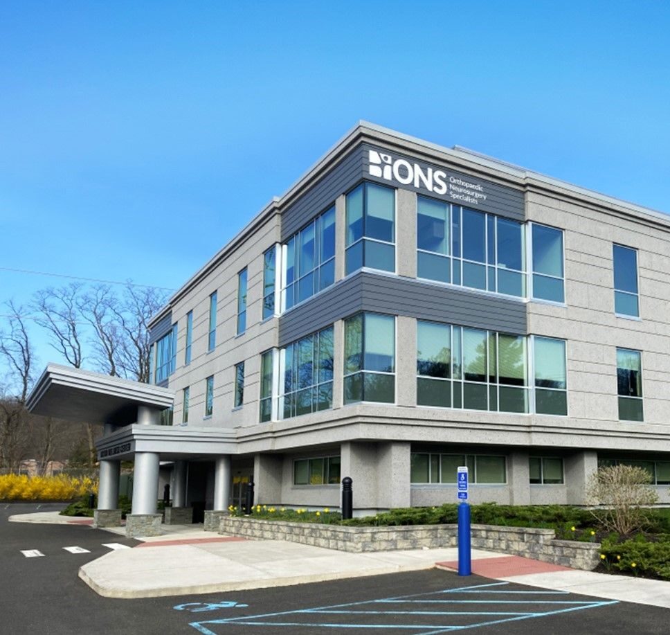
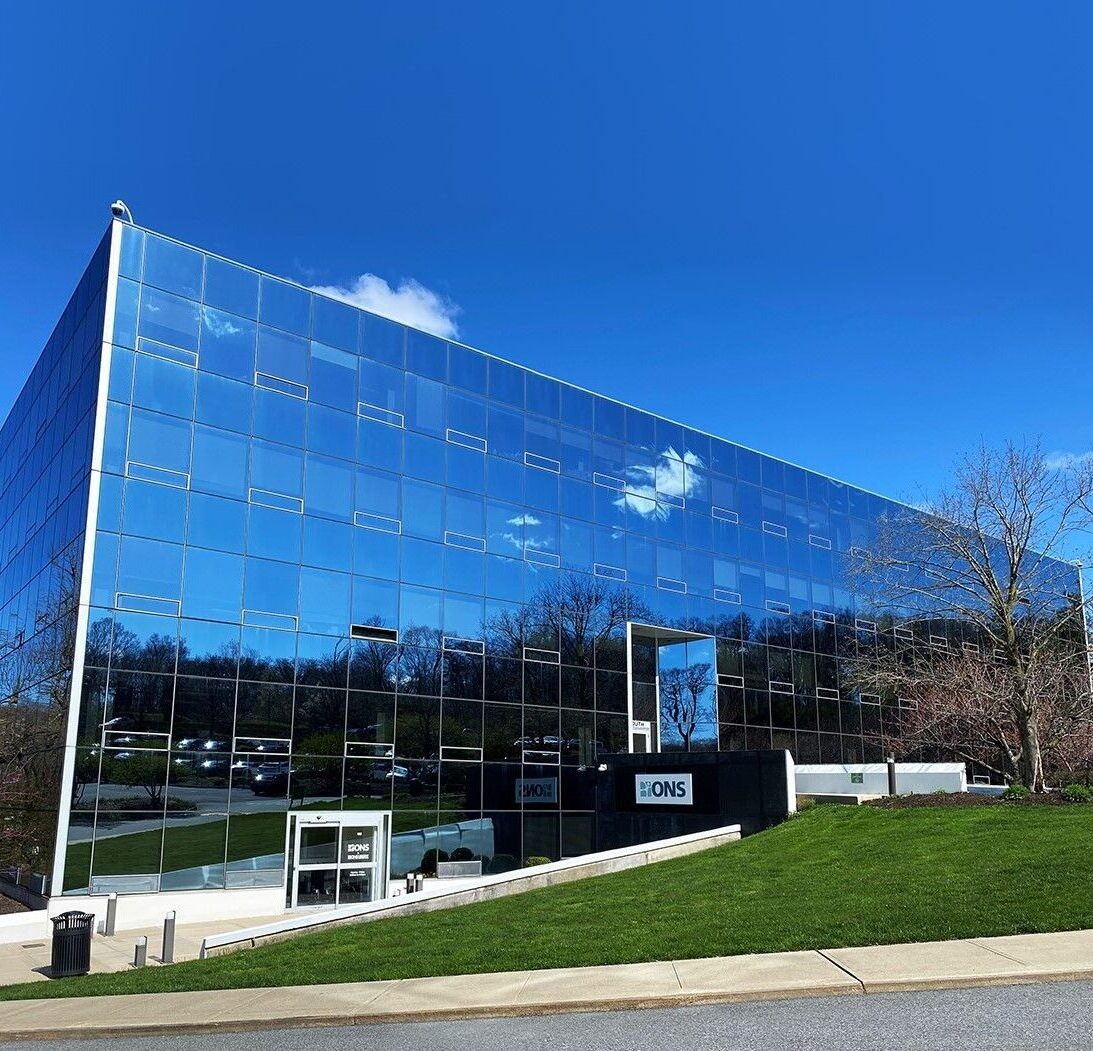
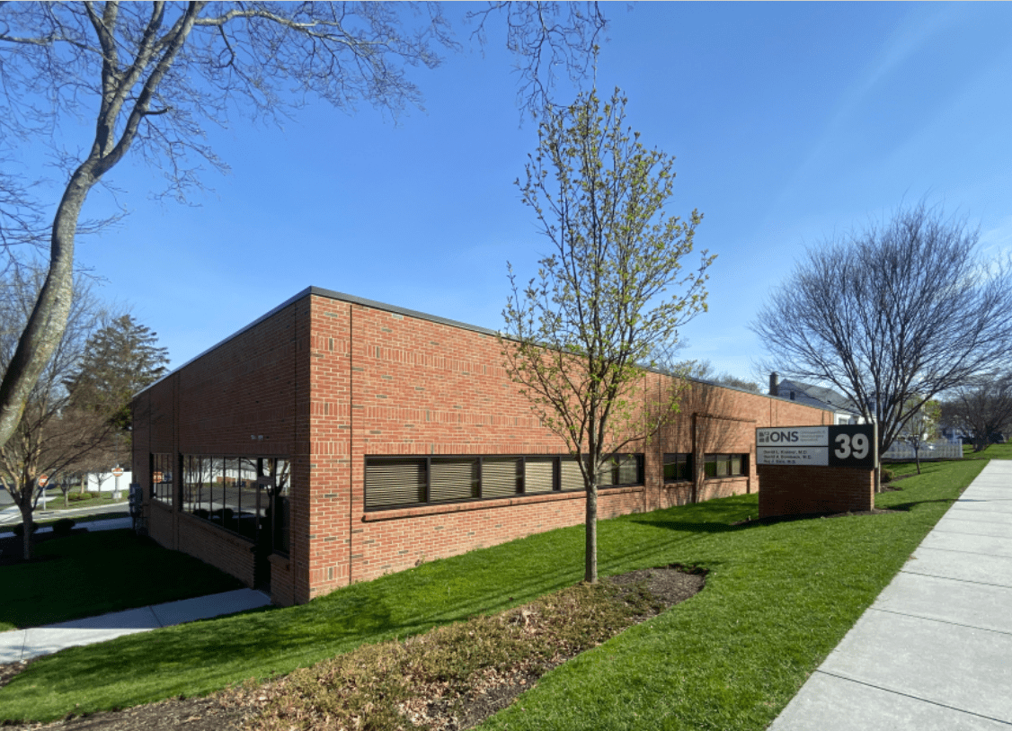
With multiple locations across the region, find the one that is most convenient for you.
You tell us where it hurts, and we take it from there
What Our Patients Say About Us
News & Blogs


Community Partners